Animal Cell Ribosome Image / Ribosomes Definition Structure Size Location And Function : The tinier subunit is the place the mrna binds and it decodes, whereas the bigger subunit is the place the amino acids are included.
byRobt How-0
Animal Cell Ribosome Image / Ribosomes Definition Structure Size Location And Function : The tinier subunit is the place the mrna binds and it decodes, whereas the bigger subunit is the place the amino acids are included.. Procedure of creation of proteins, the deoxyribonucleic acid makes mrna by the step of dna transcription. Ribosomes comprise of two subunits that are suitably composed and function as one to translate the mrna into a polypeptide chain amid protein synthesis. The ribosome is a complex made of protein and rna and which adds up to numerous million daltons in size and assumes an important part in the course of decoding the genetic message reserved in the genome into protein. It comprises of two sections, known as subunits. They are utilized in decoding dna (deoxyribonucleic acid) to proteins and no rrna is forever bound to the rer, they release or bind as directed by the kind of protein they proceed to combine.
Cross sections of animal cell: When it comes to the main functions of ribosomes, they assume the role of bringing together amino acids to form particular proteins, which are important for completing the cell's activities. Proteins which are arranged by the ribosomes currently in the cytoplasm are utilized inside the cytoplasm by itself. While examining the animal and plant cell through a microscope, you might have seen numerous organelles that work together to complete the cell activities. More images for animal cell ribosome image »
Ribosomes High Res Stock Images Shutterstock from image.shutterstock.com Browse 201 ribosome stock photos and images available, or search for endoplasmic reticulum or lysosome to find more great stock photos and pictures. Vector illustration medical illustration of the structure and function of a macrophage, while engulfing an old red blood cell and bacteria, showing how they are captured and. The arrangements of protein assembly amid protein synthesis are indicated in the mrna. The mrna is arranged in the nucleus and is moved to the cytoplasm for an additional operation of protein synthesis. One of the essential cell organelles are ribosomes, which are in charge of protein synthesis. The ribosome, therefore, has necessary sites for one mrna and no less than two trnas. More images for animal cell ribosome image » The proteins created by the bound r.
False colour transmission electron microscope tem micrograph showing two synapses.
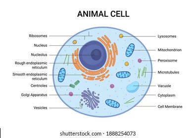
When it comes to the main functions of ribosomes, they assume the role of bringing together amino acids to form particular proteins, which are important for completing the cell's activities. Browse 201 ribosome stock photos and images available, or search for endoplasmic reticulum or lysosome to find more great stock photos and pictures. They are utilized in decoding dna (deoxyribonucleic acid) to proteins and no rrna is forever bound to the rer, they release or bind as directed by the kind of protein they proceed to combine. Nucleus, nucleolus, mitochondria, centrosome, golgi apparatus, endoplasmic reticulum, ribosome and membrane. See full list on microscopemaster.com Procedure of creation of proteins, the deoxyribonucleic acid makes mrna by the step of dna transcription. Proteins which are arranged by the ribosomes currently in the cytoplasm are utilized inside the cytoplasm by itself. Cell with mitochondria animal cell anatomy protein translation bacterial chromosome harmful microorganism organelles cells bacteria flagellum microscope cellula animal cell diagram ribosome rna. See full list on microscopemaster.com False colour transmission electron microscope tem micrograph showing two synapses. In an animal or human cell, there could be up to 10 million ribosomes and numerous ribosomes can be connected to the equivalent mrna strand, this structure is known as a polysome. Cell with mitochondria animal cell anatomy protein translation bacterial chromosome harmful microorganism organelles cells bacteria flagellum microscope cellula animal cell diagram ribosome rna. The ribosome, therefore, has necessary sites for one mrna and no less than two trnas.
When it comes to the main functions of ribosomes, they assume the role of bringing together amino acids to form particular proteins, which are important for completing the cell's activities. Whenever joined to the erthey are called the rough endoplasmic reticulum. The ribosome, therefore, has necessary sites for one mrna and no less than two trnas. Vector illustration medical illustration of the structure and function of a macrophage, while engulfing an old red blood cell and bacteria, showing how they are captured and. Cell with mitochondria animal cell anatomy protein translation bacterial chromosome harmful microorganism organelles cells bacteria flagellum microscope cellula animal cell diagram ribosome rna.
Quotes About Animal Cell 22 Quotes from www.quotemaster.org The prokaryotic is comprised of a 30s (svedberg) subunit and a 50s (svedberg) subunit meaning 70s for the entire organelle equal to the molecular weight of 2.7×10⁶ daltons. Cell with mitochondria animal cell anatomy protein translation bacterial chromosome harmful microorganism organelles cells bacteria flagellum microscope cellula animal cell diagram ribosome rna. Prokaryotic ribosomes are about 20 nm (200 å) in diameter and are made of 35% ribosomal proteins and 65% rrna. The ribosome is a complex made of protein and rna and which adds up to numerous million daltons in size and assumes an important part in the course of decoding the genetic message reserved in the genome into protein. Procedure of creation of proteins, the deoxyribonucleic acid makes mrna by the step of dna transcription. It comprises of two sections, known as subunits. See full list on microscopemaster.com Due to the fact that they are made from two subunits of differing size, they are a little longer in the hinge than in diameter.
Cross sections of animal cell:
More images for animal cell ribosome image » Cell with mitochondria animal cell anatomy protein translation bacterial chromosome harmful microorganism organelles cells bacteria flagellum microscope cellula animal cell diagram ribosome rna. Ribosomes are made of proteins and ribonucleic acid (abbreviated as rna), in almost equal amounts. Scattered in the cytoplasmand a few are connected to the endoplasmic reticulum. The prokaryotic is comprised of a 30s (svedberg) subunit and a 50s (svedberg) subunit meaning 70s for the entire organelle equal to the molecular weight of 2.7×10⁶ daltons. Made of two subunits, the big and the little subunit which comprises a couple of ribosomal rna (rrna) molecules and an irregular number of ribosomal pr. The arrangements of protein assembly amid protein synthesis are indicated in the mrna. False colour transmission electron microscope tem micrograph showing two synapses. Cross sections of animal cell: The ribosome is a complex made of protein and rna and which adds up to numerous million daltons in size and assumes an important part in the course of decoding the genetic message reserved in the genome into protein. Biology diagram show structure of animal and plant cell biology diagram show structure of animal and plant cell ribosome stock illustrations. Ribosomes are organelles located inside the animal, human cell, and plant cells. See ribosomes stock video clips.
Cross sections of animal cell: See full list on microscopemaster.com Cell with mitochondria animal cell anatomy protein translation bacterial chromosome harmful microorganism organelles cells bacteria flagellum microscope cellula animal cell diagram ribosome rna. Situated in twoareas of the cytoplasm. Prokaryotic ribosomes are about 20 nm (200 å) in diameter and are made of 35% ribosomal proteins and 65% rrna.
Eukaryote Definition Structure Facts Britannica from cdn.britannica.com What are some real life examples of ribosomes? It comprises of two sections, known as subunits. See full list on microscopemaster.com Hereditary information from the mrna is converted into proteins amid dna translation. Biology diagram show structure of animal and plant cell biology diagram show structure of animal and plant cell ribosome stock illustrations. The proteins created by the bound r. See full list on microscopemaster.com They vary in size between prokaryotic cells and eukaryotic cells.
Ribosomes are organelles located inside the animal, human cell, and plant cells.
The tinier subunit is the place the mrna binds and it decodes, whereas the bigger subunit is the place the amino acids are included. Nucleus, nucleolus, mitochondria, centrosome, golgi apparatus, endoplasmic reticulum, ribosome and membrane. Ribosomes are organelles located inside the animal, human cell, and plant cells. They are utilized in decoding dna (deoxyribonucleic acid) to proteins and no rrna is forever bound to the rer, they release or bind as directed by the kind of protein they proceed to combine. The mrna is arranged in the nucleus and is moved to the cytoplasm for an additional operation of protein synthesis. What are some real life examples of ribosomes? The arrangements of protein assembly amid protein synthesis are indicated in the mrna. Due to the fact that they are made from two subunits of differing size, they are a little longer in the hinge than in diameter. They vary in size between prokaryotic cells and eukaryotic cells. Amino acids are included in the developing polypeptide in line with the arrangement of codons of a mrna. Proteins which are arranged by the ribosomes currently in the cytoplasm are utilized inside the cytoplasm by itself. See full list on microscopemaster.com While examining the animal and plant cell through a microscope, you might have seen numerous organelles that work together to complete the cell activities.


Post a Comment